Lipomas are the most common subcutaneous mesenchymal neoplasm commonly seen over trunks and limbs. Even though vulvar lipomas are rare, their clinical nature is similar to lipomas in other sites of the body. Diagnosis is usually made on clinical findings, and surgical excision is the best treatment option. This is a case of large pedunculated vulvar lipoma in a 30-year-old lady.
vulvar lipoma, labial mass, liposarcoma
CT: computerized tomography; MRI: magnetic resonance imaging
Lipomas are a type of benign mesenchymal neoplasm that consists of normal fat cells. This is the commonest subcutaneous neoplasm in humans [1]. They are soft and fleshy on palpation. Some may grow up to 20 cm and cause disfigurement. Lipomas are commonly seen over the neck and upper back, abdomen, shoulders, buttocks, and proximal portions of the extremities [2]. Rarely it can be found in the peritoneal cavity and within the muscles. Except very few cases, vulvar lipomas are rare [3, 4]. Histological appearance and clinical presentation are almost similar to the lipomas over the other areas of the body and commonly found among young and middle-aged women [5]. Even though bilateral vulvar lipomas are rare, clinical presentation may vary from mass with ill-defined margins to pedunculated mass [3]. Diagnosis is usually made based on clinical characteristics as it is apparent on examination [6]. Even though malignant transformation has not been reported, surgical excision was done due to the risk of malignancy in the past [1]. Currently, lipomas are considered free of malignant risk [1]. This is a case of pedunculated large vulvar lipoma in a 30-year-old lady.
A 30-year-old mother of a 4-year-old child (delivered vaginally) presented with a painless pedunculated right-side vulvar mass for the last 12 years. It gradually increased in size during the first 6 years after noticing it, and there was no recent rapid enlargement afterward. Other than the mechanical difficulties due to the lump, she didn’t have any other symptoms, which made her mind not to seek medical attention. She didn’t have a family history of similar lumps. This lump was first detected by the healthcare workers at the time of her first pregnancy. She underwent normal vaginal delivery with left mediolateral episiotomy instead of right mediolateral episiotomy. She was asked to come for further evaluation and surgical excision of the lump six weeks after the child’s birth, but she did not come.
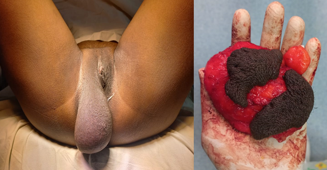
Examination revealed a soft, lobulated, non-tender, pedunculated mass (7 × 7 × 13 cm) arising from the right labia with a broad-based stalk. The overlying skin was similar to the contralateral labia majora skin. There was no regional lymphadenopathy. Her abdominal examination and pelvic examination were normal. There were no cough impulses suggestive of a hernia. The rest of the systemic examination was normal.
She underwent an ultrasound scan of the mass and groin region, which confirmed the presence of pedunculated lipoma. She was counseled regarding the diagnosis and agreed to the surgical excision.
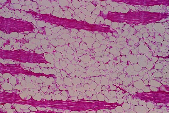
A vertical incision was made along the mass, and a lipoma was dissected out beneath the skin. The remaining skin flaps were re-fashioned and sutured. She had an uneventful recovery, and histology confirmed the diagnosis of lipoma (Figures 1 and 2).
 Figure 1: Left: vulvar lipoma before the surgery; Right: excised neoplasm with overlying skin.
 Figure 2: Histological appearance of a lipoma.
Even though it has not affected the patient in a life-threatening manner, delay in seeking treatment should be highlighted even after the detection of the neoplasm by healthcare workers. This allowed the neoplasm to grow up to a large size, which created mechanical difficulties.
Differential diagnoses in this kind of lesion can be categorized into two (benign and malignant). Benign causes are hernia, Bartholin cyst, Gartner’s cyst, lymphangioma, lipoma, sclerosing adenosis, hidradenoma papilliform, apocrine adenoma, phyllodes tumor, fibrocystic disease, and syringoma. Extramammary Paget’s disease, liposarcoma, vulvar carcinoma, ductal carcinoma, and mucinous carcinoma are the possible malignant causes [7].
Vulvar lipomas are subcutaneous, soft, multiloculated, painless, slow-growing mesenchymal neoplasms that arise from vulvar fat pads which contain fat cells interspersed with fibrous connective tissues [3, 8]. It can be seen in any age starting from infancy up to the ninth decade of life even though the exact pathophysiology has not been understood yet [9, 10]. This is more common among women than men with the highest incidence in the third and fourth decade. Even though some suggest a relationship with vulvar trauma, this patient denied any history as such [10]. All these clinical characteristics allow clinicians to diagnose this neoplasm without investigations [11]. However, it should be differentiated from liposarcoma (which has the same clinical picture), aggressive angiomyxoma, cystic swelling of the Nuck’s canal, cystic swelling of the Bartholin gland, and hernias (especially in children) [4].
Ultrasound scan is a good investigation in a low-resource setting instead of using computerized tomography (CT) scan or magnetic resonance imaging (MRI) [2]. MRI is a good tool to differentiate a lipoma from liposarcoma, as contrast-enhanced MRI shows septal enhancement in liposarcomas [4]. Other than that, both CT and MRI scans help to get a better idea regarding tumor extension and anatomical connections.
The best treatment option would be the complete surgical excision of the neoplasm, even though some suggest liposuction or steroid local injections, which are not allowed for a histological evaluation [2]. The lobular proliferation of adipocytes surrounded by thin capsules is the usual histological finding [4]. Even though recurrences can be seen after surgical excision, immediate recurrences should be carefully managed as there may be a possibility of an underlying malignant component [12].
In this patient, surgical excision is the best treatment modality as it reverts all her difficulties due to the large pedunculated mass while allowing for the histological evaluation. Other than that, it changed her vulvar disfigurement back to normal.
Vulvar lipoma is a rare condition usually diagnosed on clinical grounds. Advanced imaging may be needed in cases where the possibility of underlying malignancy needs to be excluded. Surgical excision leads to a complete cure of the disease, even though recurrences are possible.
Informed written consent was obtained from the patient.
The authors confirm sole responsibility for study conception and design, data collection, analysis and interpretation of results, and manuscript preparation.
![]() *1, Wickramasinghe NH2 and Senevirathne JTN2
*1, Wickramasinghe NH2 and Senevirathne JTN2

