The Misleading Ocular Squamous Lesions

Sebbata SORCID Logo*1, Brarou HORCID Logo1, Laaouina SORCID Logo1, Khachani K2, Oubaaz B1, Fiqhi A1, Abdellaoui TORCID Logo3, Mouzari Y1 and Oubaaz A1

1Department of Ophthalmology, Mohammed V Military Hospital, Rabat, Morocco
2Department of Dermatology, Ibn Sina Hospital, Rabat, Morocco
3Mohamed Ben Abdellah University, Fez, Morocco

*Corresponding author: Soundouss Sebbata, Department of Ophthalmology, Mohammed V Military Hospital, Rabat, Morocco

Received: 16 November 2023; Accepted: 29 December 2023; Published: 01 January 2024

© 2024 The Authors. This is an open-access article and is distributed under the Creative Commons Attribution License, which permits unrestricted use, distribution, and reproduction in any medium provided the original work is properly cited.

Keywords

lesions, ophthalmology, biomicroscopy, psoriasis, dermatologists

Clinical Image

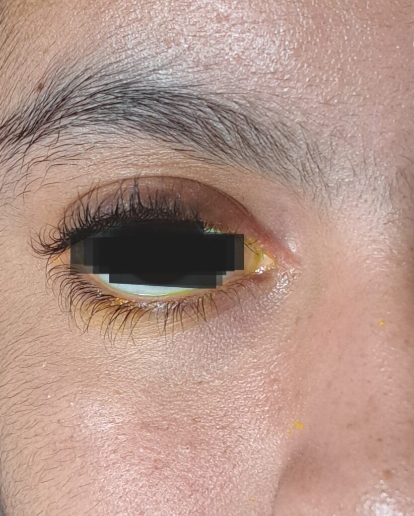
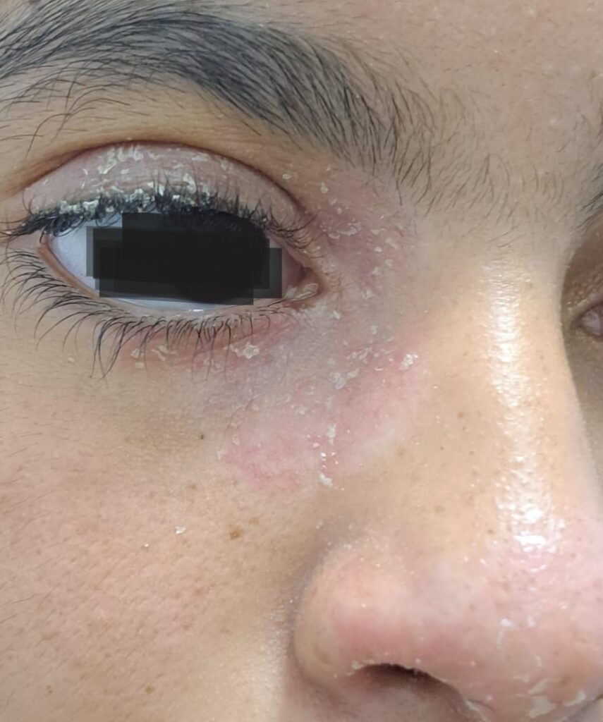
Figure 1: Patient with unilateral blepharitis associated with dermal squamous lesions on an erythematous base, affecting the palpebra and the nose.

Figure 2: Improvement in the clinical condition after two months of treatment.

A 14-year-old girl presented to the ophthalmology department for unilateral blepharitis associated with dermal squamous lesions on an erythematous base, affecting the palpebra and the nose (Figure 1). There was no visual impairment or any other ocular sign. On biomicroscopy, there was no tear film damage. The break-up time was counted at 12 seconds. An initial treatment based on good lid hygiene was prescript, and the patient was referred to the dermatologist for suspected eczema. Later on, the same lesions were also noted in the palmoplantar area. After the failure of an initial treatment, a biopsy of the lesions was performed, revealing psoriasis affection. A lubricant ointment was added, with multiple applications per day. We noticed an improvement in the clinical condition after two months (Figure 2). No topical corticosteroid was used.

Psoriasis is a persistent skin condition that can affect any part of the body. Occasionally, its most unusual localizations involve atypical anatomic locations. Facial involvement is estimated to occur only in 17–46% of patients [1]. Palpebral affection is extremely rare, even in the erythrodermic variant of the disease. Uveitis is more common but was not found in our patient [2].  Affection of the eyelids leads to challenging treatment issues, as the application of steroids is prohibited by some dermatologists due to the local side effects. They suggest that eyelids should never be without a thin film of a moisturizing cream [3], hence our therapeutic approach. Our analysis crystallizes the fact that close collaboration between ophthalmologists and dermatologists is essential in the great management of ocular-dermatologic lesions.

References