Akanksha*1, Govil V1, Yadav D2, Kumar TH1, Kumar S1 and Singh A3
1Department of Anaesthesiology, Pt. B.D. Sharma PGIMS, Rohtak, Haryana, India
2Department of Radiodiagnosis, Pt. B.D. Sharma PGIMS, Rohtak, Haryana, India
3Kasturba Medical College, Mangalore, India
*Corresponding author: Dr. Akanksha, Department of Anaesthesiology, Pt. B.D. Sharma PGIMS, Rohtak, Haryana, India
Received: 29 November 2023; Accepted: 01 January 2024; Published: 08 January 2024
© 2024 The Authors. This is an open-access article and is distributed under the Creative Commons Attribution License, which permits unrestricted use, distribution, and reproduction in any medium provided the original work is properly cited.
Abstract
Introduction: Patients with multiple myeloma (MM) have improved life expectancy and quality of life in the last decade with the introduction of new chemotherapeutic agents and improvement in treatment modalities. Due to this, patients are now being treated for surgical implications of the disease, which was not possible earlier.
Case Report: We report a case of a 42-year male with MM with fractured femur bone posted for fixation of pathological fracture femur, discussing the perioperative considerations and management for anaesthesiologists.
Conclusion: Proper anaesthetic management is crucial for the successful fixation of pathological fractures in patients with MM and comorbidities, considering the unique challenges posed by the disease and its associated complications.
Keywords
anaesthesia, multiple myeloma, pathological fracture
Abbreviations
MM: multiple myeloma, IV: intravenous, OETT: oro-endotracheal tube
1. Introduction
Of all hematological malignancies, multiple myeloma (MM) has an incidence of 13% [1]. With the introduction of new chemotherapeutic regimens for MM, the quality of life and survival rates have improved in these patients. The mean 5-year- survival rate of MM has increased from 25% in the 1970s to >50% in the last decade [2]. Therefore, now orthopedic surgeries are possible for patients with MM for treatment of pathological fractures. Due to the paucity of literature regarding the anaesthetic management of these patients, we would like to highlight the pathophysiology and the clinical symptomatology of this disease, the side effects of the chemotherapeutic agents, and emphasize the guidelines to continue the appropriate supportive therapy during anaesthetic management in the perioperative period.
2. Case Report
An obese 42-year-old man, diabetic and hypertensive, weighing 110 kg with a BMI of 33.1 kg/m2, presented to our hospital with a history of pain in the left hip from the last 3 months. On detailed history, the patient revealed that he usually snored during sleep and regularly woke because of apnea, and this symptom was more marked in the supine position; therefore, he normally slept in a lateral position. The patient was diagnosed with obstructive sleep apnea syndrome (OSAS). A physical examination performed on admission showed that his neck was short and stiff, with a restriction of activity and poor neck mobility, and his mouth could only be opened to the width of two and a half horizontal fingers (Figures 1–4). An airway assessment was performed, and the patient’s Mallampati score was 3, thyromental distance (TMD) was 2.5 cm, and the upper lip bite test was 3.
The patient was taking a tablet of metformin 1000 mg/day per oral (PO) for diabetes and a tablet of amlodipine 10 mg/day per oral (PO) for hypertension and was controlled on these medications. He was diagnosed with MM 2 months ago, for which he underwent 4 cycles of chemotherapy with injection bortezomib 1.3 mg/m2 subcutaneously. X-ray of the hip showed a pathological fracture of the left neck of the femur (Figure 5). He was taking a tablet of lenalidomide 20 mg/day per oral (PO), a tablet of dexamethasone 40 mg/week per oral (PO), a tablet of aspirin 75 mg once daily (OD) orally, and a tablet of atorvastatin 20 mg once daily (OD) orally for antithrombotic prophylaxis. Blood investigation showed normal blood counts with haemoglobin – 14.1 gm%, total leucocyte count – 11,000, platelet count – 1.72 lakh, and normal renal function with blood urea – 28 mg/dL, serum creatinine – 0.6 mg/dL, serum sodium – 134 mEq/L, serum potassium – 4.68 mEq/L. Urine complete examination was normal, revealing no ketone bodies and albuminuria. Serum calcium was 8.3 mg/dL, and serum albumin was 3.2 g/dL. Glycated haemoglobin was 5.6, and random blood sugar was 184 mg/dl revealing adequate blood glucose control. The coagulation profile of the patient was deranged with PT 36.6 seconds and INR of 2.7, which was optimized with a transfusion of 4 units of fresh frozen plasma. The electrocardiogram and chest X-ray were normal. USG color doppler for the left lower limb was done to rule out any venous thrombosis. It was decided to continue aspirin, and preoperative antibiotic prophylaxis was administered with an injection of cefoperazone + salbactum 1.5 g intravenous (IV). A wide-bore intravenous cannula was secured, and the patient was hydrated with 10 mL/kg of normal saline before induction of anaesthesia. Anaesthesia was induced with an injection of fentanyl 120 mcg IV, an injection of propofol 140 mg IV, an injection of atracurium 40 mg IV, and the airway was secured with tracheal intubation with an oro-endotracheal tube (OETT) size 8 mm internal diameter. Under all aseptic precautions, left radial artery cannulation was done to monitor invasive BP and samples for arterial blood gas analysis were sent. Anaesthesia was maintained with FiO2 0.5 (air oxygen mixture) and sevoflurane maintaining 0.8–1.2 MAC. After induction, the patient was positioned in the right lateral for surgery to be started with adequate padding of all pressure points. The procedure was uneventful, with no hemodynamic disturbance and minimal/allowable blood loss. After surgery, inhalation anaesthetics were stopped, and the OETT was removed after reversing the patient with an injection of glycopyrrolate 0.6 mg IV and an injection of neostigmine 3.5 mg IV. The patient had an uneventful recovery. Postoperatively, the patient was comfortable. He did not require any rescue analgesics. Renal function tests (blood urea – 38 mg/dL, serum creatinine – 0.8 mg/dL) and serum calcium (8.3 mg/dL) were normal in the postoperative period. He was discharged on the fifth postoperative day (Figure 6).
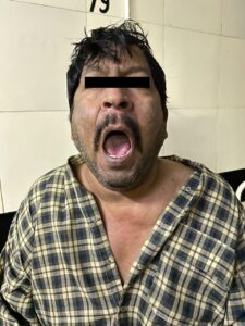
Figure 1: Patient with adequate mouth opening. Figure 2: Patient with tongue protruding.
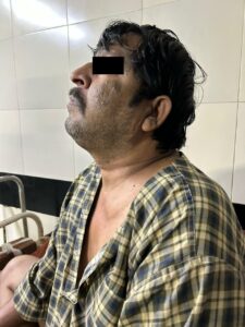
Figure 3: Patient with limited extension.
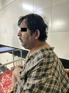 Figure 4: Patient with limited flexion.
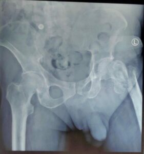 Figure 5: X-ray of the left femur showing pathological fracture of the neck of the femur.
 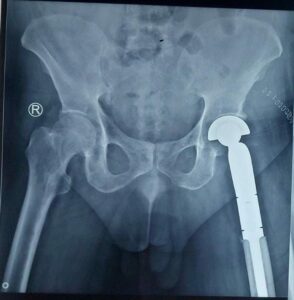 Figure 6: X-ray of the left femur showing mega prosthesis in situ postoperatively.
3. Discussion
MM, also known as Kahler’s disease, is a neoplastic proliferation of plasma cells in bone marrow derived from a single clone. MM is the malignant proliferation of monoclonal plasma cells in the bone marrow, leading to increased production of abnormal antibodies and secretion of monoclonal immunoglobulins detectable in the blood or urine and associated with organ dysfunction. The clinical manifestations of symptomatic MM are lytic or osteopaenic bone lesions, hypercalcaemia, renal failure, anaemia, and infections, but patients are now diagnosed earlier at an asymptomatic stage. Due to multi-system involvement, it is important to optimize these patients preoperatively for a better postoperative outcome. Renal dysfunction is a common complication of MM. Renal insufficiency is seen in nearly one-half of MM patients at presentations and is associated with increased mortality [3]. Renal failure commonly occurs because of damage to the renal tubules by the free light chains and hypercalcaemia due to osteolysis. Adequate hydration, correction of hypercalcaemia and hyperuricemia, and antimyeloma therapy should be initiated promptly preoperatively to improve postoperative outcomes. Although the renal function tests were normal in our patient, we ensured adequate hydration by preloading the patient with IV fluids, maintaining the haemodynamic, and avoiding nonsteroidal anti-inflammatory drugs (NSAIDs) by other adjuvants. The patient’s serum calcium levels were monitored in the perioperative period. Adequate hydration prevents hypercalcaemia. Severe hypercalcaemia should be treated with thiazide and bisphosphonates [4, 5]. Preventing and reversing renal failure in patients with MM improves long-term survival [6]. Also, increased plasma cell proliferation depletes the normal stromal cells of the marrow, leading to anaemia and thrombocytopenia, rendering these patients at high risk for excess bleeding intraoperatively, requiring transfusion of blood and platelets. Target haemoglobin of 12 g/dl preoperatively is recommended in these patients [7]. Patients with MM have increased susceptibility to infection, lytic lesions in bone, renal failure, hemorrhagic tendencies, hyperviscosity syndrome, anaemia, and hypercalcaemia. Apart from these, patients also develop adverse events related to their drug therapy. Newer drugs such as thalidomide, lenalidomide, and bortezomib have prolonged survival rates but have serious adverse effects as well. Haematological toxic effects such as neutropenia and thrombocytopenia are common with these drugs when used along with conventional chemotherapy. The incidence of venous and arterial thrombosis increases with thalidomide and lenalidomide. Since our patient was on lenalidomide, he was started on low-dose aspirin for thromboprophylaxis. As per the International Myeloma Working Group guidelines, the patient had >2 risk factors for thromboembolism (lenalidomide therapy, high-dose steroids, diabetes, surgery, and anaesthesia administration) therefore, he was continued with aspirin for thromboprophylaxis. Hypotension in the perioperative period, along with increased risk for thromboembolism, puts these patients at high risk for stroke and myocardial ischaemia (MI) [8]. Aspirin was continued after surgery, and thromboembolism deterrent compression stockings were applied in the lower limbs to prevent deep vein thrombosis [9, 10]. These patients are more prone to infections due to neutropenia and immunosuppressive drugs. A prophylactic antibiotic was administered for our patient, and absolute asepsis was maintained.
4. Conclusion
Many MM patients are now being anaesthetised for orthopaedic procedures in the last decade, with improved survival rates after the introduction of new chemotherapeutic agents. Specific guidelines are available for the supportive therapy of these patients to prevent renal failure, infections, anaemia, and thromboembolism. As anaesthesiologists, we must know these guidelines to prevent long-term complications when these patients are under our care for a short perioperative period.
Clinical Message
Cautious selection of anaesthesia techniques and close collaboration between the surgical and anaesthetic teams are crucial for the successful fixation of pathological fractures in patients with MM and comorbidities, prioritizing optimal pain control, minimizing perioperative complications, and ensuring favorable patient outcomes.
Consent for Publication
Consent of the patient has been taken so that his photo and details can be published in the case report.
Funding
No funding was available.
Conflicts of Interest
The authors declare that they have no conflict of interest.
References
- Palumbo A, Anderson K. Multiple myeloma. N Engl J Med. 2011;364(11):1046-060.
- National Institutes of Health. SEER Relative Survival (Percent) By Year of Diagnosis, All Races, Males and Females.
- Heher EC, Rennke HG, Laubach JP, et al. Kidney disease and multiple myeloma. Clin J Am Soc Nephrol. 2013;8(11):2007-017.
- Park KJ, Menendez ME, Mears SC, et al. Patients With Multiple Myeloma Have More Complications After Surgical Treatment of Hip Fracture. Geriatr Orthop Surg Rehabil. 2016;7(3):158-62.
- Ludwig H, Zojer N. Supportive therapy in multiple myeloma. Recent Results Cancer Res. 2011;183:307-33.
- Bisoyi S, Pratihary BN, Mohapatra R, et al. Perioperative considerations in the management of a patient with multiple myeloma undergoing aortic valve replacement. J Cardiothorac Vasc Anesth. 2015;29(1):151-55.
- Terpos E, Kleber M, Engelhardt M, et al. European Myeloma Network. European Myeloma Network guidelines for the management of multiple myeloma-related complications. Haematologica. 2015;100(10):1254-266.
- Wang CJ, Cheng KI, Soo LY, et al. Intraoperative stroke under epidural anesthesia for bipolar hemiarthroplasty in a patient with multiple myeloma: a case report. Kaohsiung J Med Sci. 2001;17(1):55-9.
- Schoen MW, Carson KR, Luo S, et al. Venous thromboembolism in multiple myeloma is associated with increased mortality. Res Pract Thromb Haemost. 2020;4(7):1203-210.
- Cesarman-Maus G, Braggio E, Fonseca R. Thrombosis in multiple myeloma (MM). Hematology. 2012;17 Suppl 1(0 1):S177-80.


