Abstract
Morel-Lavallée lesion (MLL) is a closed degloving soft tissue injury that occurs when there is abrupt separation of skin and subcutaneous tissue from the underlying fascia. It usually develops following high-velocity trauma and crush injuries. This is a case report of a young female who had a fall and presented with a large swelling in her left thigh and was diagnosed to have a MLL. She underwent drainage of the collection; the wound was left open and allowed to heal by secondary intention.
Keywords
Morel-Lavallée lesion, thigh swelling, thigh abscess, incision and drainage
Abbreviations
MLL: Morel-Lavallée lesion
1. Introduction
The Morel-Lavallée lesion (MLL) was first described by the French physician Maurice Morel-Lavallée in 1853 as a closed degloving injury in the pelvis. A shearing force results in skin and subcutaneous tissue being separated from the underlying fascia. Disruption of bridging blood and lymphatic vessels leads to a collection of fluid in the plane created as a result of the injury [1].
This fluid is composed of blood, lymph, and necrotic fat. Granulation tissue forms around this collection and may become organized into a fibrous pseudocapsule. The presence of such a capsule prevents absorption of the contained fluid, leading to a gradual expansion of the collection. This explains why the clinical presentation of these lesions can be several months or even years following injury [2].
Development of MLL is usually seen following high-velocity trauma and crush injuries. They are often associated with fractures of the acetabulum and pelvis [3].
This report highlights a case of a high-volume MLL of the left thigh in a 27-year-old Indian woman.
2. Case Report
A 27-year-old female presented to our OPD with complaints of swelling in the lateral aspect of her left thigh. She first noticed the swelling a fortnight before she presented to our center, and she claimed that it had increased to double its initial size during the period.
On further probing, she gave us a history of trauma to the lateral aspect of the left thigh around 6 months back when she fell from her vehicle. She had no other history of note.
She was evaluated at a primary care center and suspected to have a thigh hematoma and was referred to our center for further care.
On examination, she looked well and walked with a normal gait.
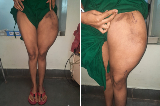
She had a large area of fullness of approximately 30 × 15 cm over the lateral aspect of her thigh, which was fluctuant and thus suggestive of a large fluid collection. There was no redness or the local rise of temperature over the swelling, and the area was non-tender (Figure 1).
 Figure 1: Fullness noted over lateral aspect of left thigh.
A contrast-enhanced CT scan of the thigh was then done, which illustrated a large, well-defined fusiform encapsulated hypoattenuating collection of 9 × 7 × 30 cm with a volume of 1100cc in the deep subcutaneous plane of the left thigh extending from the level of the greater trochanter to 10 cm above the knee joint. The collection is seen separately from muscular planes. The rest of the scan was normal. The diagnosis of MLL was considered based on the history, clinical findings, and CT report.
Aspiration was performed, which revealed a clear, serous, and non-foul-smelling straw-colored fluid. Fluid analysis revealed a lymphocyte-rich collection and the fluid culture and sensitivity have no growth, further strengthening the suspicion of MLL.
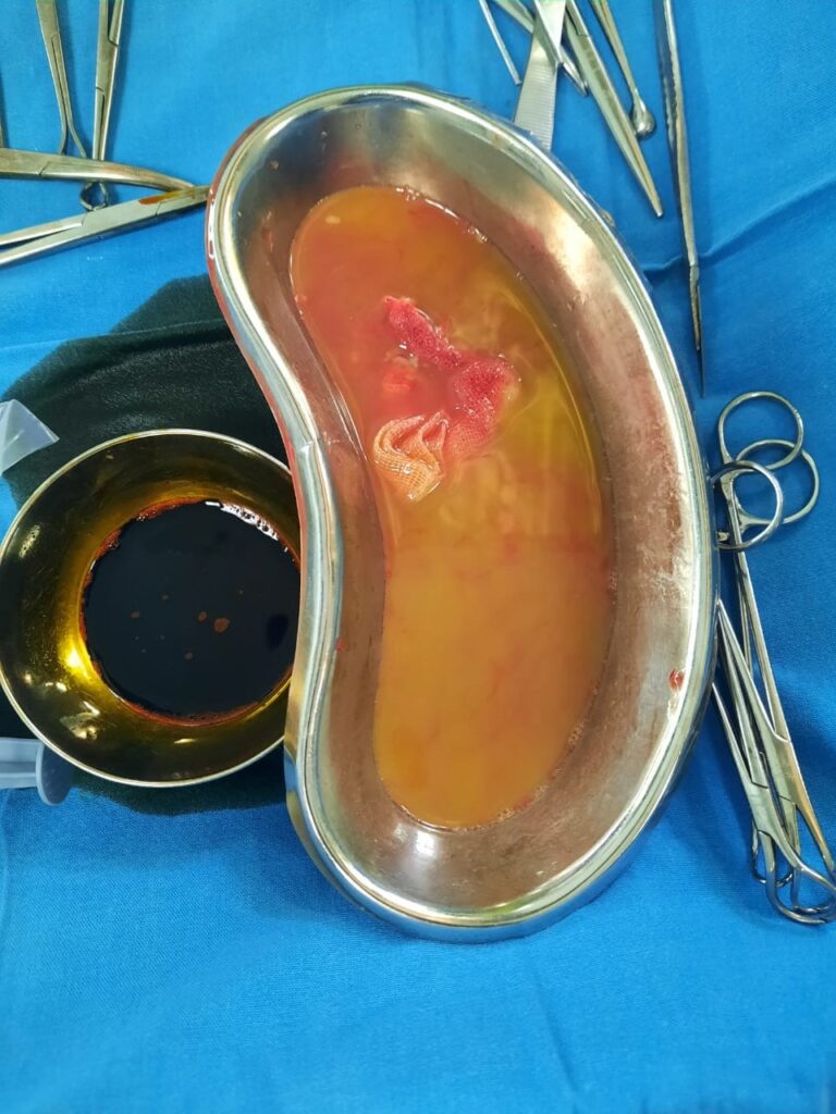
The patient underwent incision and drainage under spinal anesthesia. Intraoperatively, a capsulated collection of 1200 ml of yellowish, serous, and non-foul-smelling fluid was noted. The fluid was completely drained, and the cavity was debrided. The surgical wound was left open to allow healing by secondary intention (Figure 2).
The patient was discharged the next day, and the wound had completely healed by a month since surgery. The patient was followed up for a period of one year, and there was no recurrence.
 Figure 2: Serous, yellow fluid drained from the swelling.
3. Discussion
Although MLL were described over 150 years ago, there have been only approximately 200 reports in the literature using our search method. Their presence has significantly higher surgical site infection, so their detection is of great importance.
Though MLL is reported to be a rare entity, it is believed to have a higher incidence than the actual documented incidence, as it is often misdiagnosed and underreported.
Thus, there is a lack of sufficient statistical data and a lack of an established treatment protocol for MLL.
Key features of the history and examinations contributed to the diagnosis of MLL in this case. Due to the high rate of recurrence associated with needle aspirations of MLL, we chose surgery as our primary treatment modality.
Surgery involves evacuation of the haemolymphatic collection with excision of the pseudocapsule and debridement of necrotic tissue. The wound may be left open with or without the assistance of a vacuum dressing or closed primarily with or without a drain insitu. In this case, we have left the wound open.
The paper by Demirel et al. [4] advocates the use of synthetic glue to close the dead space intraoperatively. Their series of 7 thigh MLLs sustained from road traffic accidents (RTAs) all had a successful outcome when surgical drainage was combined with the use of synthetic glue and compression bandaging.
When misdiagnosed or in case of delayed diagnosis, MLL can result in various complications, which include secondary infections and necrosis of overlying skin. Thus, accurate diagnosis and early treatment are of great importance in the case of MLL.
4. Conclusion
MLL is a fairly uncommon entity, which can result in various complications that could even be life or limb-threatening. Thus, it is important for all healthcare workers, in particular surgeons, orthopedics, and primary care physicians, to be aware of this entity, its complications, and treatment options.
References
![]() , Sultana S and Varma V*
, Sultana S and Varma V*

