The consistency of the myxoma is often dense fibrous, or jelly-like mucous. Myxoma consists of undifferentiated mesenchymal cells of various shapes (rounded, stellate), which are located in the myxoid stroma. Myxoma is the most common primary cardiac tumor (up to 50% of all primary cardiac neoplasms). Myxoma of the stomach is a rare pathology. We could not find any information covering the issues of gastric myxoma in children. This article provides a clinical observation of a good result of surgical treatment of gastric myxoma in a 2-year-old child. The described clinical case indicates the need to create a unified register of rare tumors for their study.
myxoma, tumor, children, stomach
PAMT: plexiform angiomyxoid myofibroblastic tumor, WHO: World Health Organization
Myxoma is a benign tumor composed of spindle-shaped cells surrounded by myxoid tissue. Myxomas are commonly found in the heart, skin, and ovaries, around joints, inside nerves, and inside skeletal muscles. Myxoma is a benign (non-cancerous) type of tumor [1].
Myxoma is the most common primary cardiac tumor (up to 50% of all primary cardiac neoplasms) [2]. Gastric myxoma is a rare pathology [3]. At the beginning of the 21st century, Takahashi et al. [4] first described a rare mesenchymal tumor disease, plexiform angiomyxoid myofibroblastic tumor (PAMT). The neoplasm is also known as plexiform fibromyxoma and is recognized as a separate neoplasm among benign tumors of the stomach according to the WHO classification of tumors of the digestive system.
In 2016, the literature provided information on 59 morphologically verified cases of PAMT [5]; by 2019, the total number of patients with this pathology reached 113 [6], and in 2020, isolated cases of the disease were additionally described [7].
All results obtained in the course of the studies are based on the treatment of adult patients. Tumors of the stomach in children are extremely rare [8]. In the UK, in children under 14 years of age, it occurs at a frequency of 0.02 per 1 million per year [9]. In the English-language literature, as a rule, isolated cases are presented. We could not find any information covering the issues of gastric myxoma in children.
The article presents a rare clinical observation of gastric myxoma, which was first regarded as a cystic formation of the pancreas.
Patient A, at the age of 1 year 10 months, applied to the Samarkand Regional Children’s Medical Multidisciplinary Center with complaints, according to his mother, of a volumetric formation in the abdominal cavity, weakness, and lack of appetite.
From the anamnesis, it was revealed that about 1 month before contacting us, the child’s mother found a tumor-like formation in the abdominal cavity. The patient was examined in the Samarkand branch of the Specialized Scientific and Practical Medical Center of Oncology and Radiology. Ultrasound of the abdominal organs in the left half of the abdomen revealed a hypoechoic formation with smooth edges measuring 10.6 × 7.4 cm. On the computed tomography of the abdominal organs on the left, a formation with clear boundaries measuring 7.7 × 8.8 × 9.6 cm is detected (Figure 1).
 Figure 1: Multispiral computed tomography of patient A.
Based on the results of the examination, the child was diagnosed with “cystic formation of the body and tail of the pancreas.”
After the standard planned preoperative preparation on 06/09/2023 in the Samarkand Regional Children’s Medical Multidisciplinary Center with a preliminary clinical diagnosis of “dermoid cyst of the abdominal cavity”; the patient underwent the operation laparotomy, removal of mass formation of the abdominal cavity.
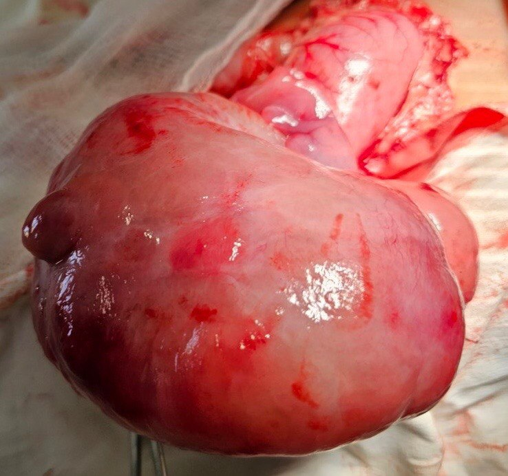
Intraoperatively, a pink-whitish mass was detected, the tumor was round in shape, 12 cm in diameter (Figure 2).
 Figure 2: Intraoperative picture of the tumor.
The boundaries of the formation were clear and not soldered to any organ of the abdominal cavity. The tumor was a dense fibrous tissue. Palpation was smooth, hard elastic consistency. The vascular pedicle of the formation emanated as a cord-like commissure from the greater curvature of the stomach (Figure 3).
 Figure 3: Macro-operator. Removed tumor.
The postoperative period proceeded smoothly. The child was discharged on the 5th day after the operation.
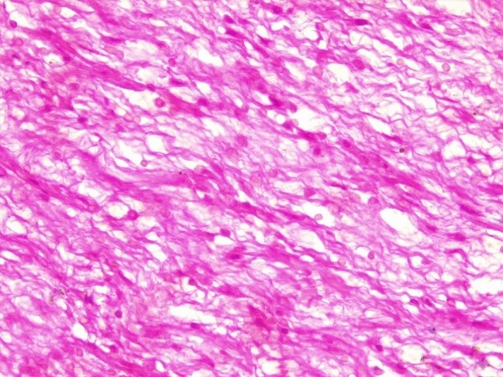
According to postoperative histopathological examination No. 1957/23 (June 24, 2023), the tumor is represented by stratified squamous epithelium, under which fibrosis- angiomatosis is noted. Myxoid tissue is founded in some regions. The stroma is composed of loose fibrous tissue with collagenization. The hypochromic nuclei of the stroma are large and eccentrically located. Conclusion: embryonic myxoma.
To prepare histological preparations, the samples were fixed in 10% neutral buffered formalin, dehydrated in ascending alcohols, and embedded in paraffin blocks, from which sections 4 μm thick were prepared. Histological sections were stained with hematoxylin and eosin (Figure 4).
 Figure 4: Microphoto myxomas of the stomach. 400 times magnification.
This clinical observation indicates the complexity of the differential diagnosis of abdominal tumors in young children. Due to the fact that gastric myxoma is a rare pathology, the leading role in the final diagnosis is given to morphological examination. Pediatric surgeons should be aware of this rare pathology and, if possible, refer patients with abdominal tumors to specialized institutions to develop an adequate treatment strategy.
![]() *1, Azizov MK2, Ulmasov FG3, Allazov F3, Tursunov SE2 and Mavlyanov ShKh1
*1, Azizov MK2, Ulmasov FG3, Allazov F3, Tursunov SE2 and Mavlyanov ShKh1


